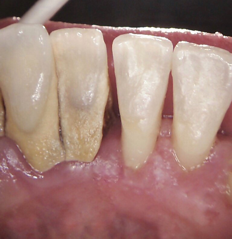
Calculus removal at Courtney dental7/6/2021 This photo is of the inside surface of lower teeth in a patient Nic recently treated over 2 appointments. The right side of the photo was cleaned 1 week prior to the photo and the left was cleaned after the photo was taken. This is a great example to show of calculus removal, you can see the gums on the left is red, inflamed and when touched bleeds easily. The gum on the right has a healthier appearance, the gum is nice and pink and their is no bleeding when probed. Calculus, sometimes called tarter, is the result of plaque build-up that hardens (calcifies) on the teeth. Once you brush your teeth, plaque begins to form on your clean teeth right away and within two to three days, the calcification process begins in the plaque and starts to turn into calculus if not removed by brushing and flossing.
Calculus has a hardened surface and provides more surface area on the tooth for plaque to adhere to. More plaque could lead to more gum and periodontal disease, so removing calculus is an important step to long-term oral health. This photo is an example of supragingival calculus which forms above the gumline; it's yellow or tan in colour and is visible on a tooth's surface. Subgingival calculus forms on the tooth root below the gumline in the sulcus (crevice) between the tooth and the gum. It typically isn't visible with the naked eye because it is hidden under the gum. Subgingival calculus is typically brown or black in colour. Once calculus collects on your teeth, it needs to be removed using a process known as debridement. Nic used hand-held instruments and an ultrasonic device to remove the calculus. The ultrasonic device incorporates a combination of high-frequency vibrations with water to remove the deposits. Comments are closed.
|
Archives
February 2024
Categories
All
|


